비중격에 발생한 근상피종 1례
Myoepithelioma of the Nasal Septum: A Case Report
Article information
Abstract
= Abstract =
Myoepithelioma is a rare benign neoplasm that mostly arises in the major salivary glands and sometimes in the minor salivary glands, which account only for less than 1% of all salivary glands neoplasms. However, its extra-salivary involvement is even rarer and only a few cases of nasal cavity myoepithelioma were reported in the English-language literature so far. In this case report, we present a 40-year-old female with unilateral nasal obstruction diagnosed as myoepithelioma of the nasal septum and treated with endoscopic sinus surgery.
서론
근상피종은 주로 대타액선 혹은 때때로 부타액선에 발생하는, 타액선 종양에서도 1% 미만의 발생 빈도를 차지하는 드문 종양이다. 타액선 외 침범은 더욱 드물게 있으며, 그 중 비강 침범은 현재까지도 불과 수 케이스 정도만이 보고될 정도로 희귀한 질환이며, 그 중 비중격에서 기원한 경우는 더욱 드물게 보고되었다.1-13) 저자들은 일측성 코막힘을 주소로 내원한 40세 여환의 비중격에서 기원한 근상피종 및 이를 내시경부비동수술로 시행한 증례에 대해 진단 및 치료적 측면과 함께 보고하고자 한다.
증례
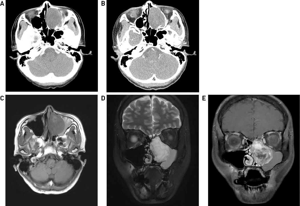
40세 여자 환자가 내원 1개월 전 우연히 자녀의 소아청소년과 의원 진료에 동반하였다가 본인의 코막힘에 대해 비강 검진을 시행 받은 뒤 좌측 비폴립 소견을 듣고 큰 병원 진료를 권유 받아 본원 이비인후과로 내원하였다. 환자는 오래전부터 지속된 좌측 코막힘을 호소하였고, 과거력상 특이 사항은 없었다. 시행한 내시경 검진상 좌측 비중격에서 기원한 것으로 추정되는 좌측 비강의 종괴가 관찰되며 이로 인한 우측으로의 비중격 만곡증 소견이 관찰되었으나 외비 변형은 없었고(Fig. 1A), 음향비강통기도검사상 비점막수축제 분무 전후로 비공으로부터 5 cm까지의 비강 부피는 우측 5.35 cm3 및 6.65 cm3, 좌측 3.20 cm3 및 3.87 cm3로 측정되었고, 제 2 절흔 부위의 비강단면적은 우측 0.61 cm 및 0.87 cm3, 좌측 0.34 cm3 및 0.55 cm3로 측정되어 모두 좌측이 작은 값을 나타내었다. 조영제를 사용한 부비동 전산화단층촬영상 좌측 비강과 사골동을 가득 채우는 4.5 cm 크기의 팽창성 병변이 관찰되었고, 이로 인해 좌측 상악동은 외측으로 전위되어 있는 소견을 보였으며, 조영 전 균질 저음영과 조영 후 지연기 비균질 조영 증강 소견이 관찰되었다(Fig. 2). 진단을 위해 부비동 자기공명영상촬영과 내시경 조직검사를 추가로 시행하였고, 조영제를 사용한 부비동 자기공명영상촬영상 병변은 조영 전 비균질 T1WI 저신호강도, T2WI 비균질 고신호강도 및 조영 후 비균질 조영 증강을 보여 소타액선종양 혹은 신경초종과 같은 양성종양이 의심되었고(Fig. 2), 조직검사 결과 근상피종으로 진단되었다. 이에 전신마취 하 좌측 내시경부비동수술을 통해 내시경적 종양적출술과 함께 접협동개방술, 사골동절제술, 중비도개창술을 시행하였다.

Pre-operative endoscopic finding shows polypoid mass (arrow) originated from nasal septum (A). After resection of tumor, thinning of middule turbinate is observed (B). Endoscopic finding after 12 month from operation shows no evidence of residual tumor or recurrence (C). S ; nasal septum, M ; middle turbintate.

Pre (A) and Post (B) contrast-enhanced PNS CT scan shows well-marginated expansile soft-tissue mass inducing pressure remodeling to adjacent bone. PNS MRI image shows T1 low-signal intensity (C), heterogeneous T2 high-signal intensity (D) and heterogeneous enhancement pattern (E) which suggest for minor salivary gland tumor or schwannoma.
네비게이션 유도 하에 수술을 시행하였고, 종양의 기원은 비중격으로 관찰되었으며, 비중격에서 종양으로부터 앞쪽으로 2 mm 경계를 두고 canal knife를 이용해 절개를 넣은 후, 비중격점막을 박리하여 기원 부위를 포함하여 종양을 완전히 적출하였다. 종양 내부는 가피와 노란색의 조직파편들로 이루어져 있었으며, 종양 제거 후 관찰 시 구상돌기와 하비갑개는 후외측으로, 중비갑개는 얇게 찌그러진 채로 상측으로 밀려 있었으며(Fig. 1B), 두개저 및 다른 부비동으로의 부착이나 종양의 잔해가 없는 것을 확인 후 surgicel을 이용하여 비강 내 패킹 시행한 뒤 수술을 종료하였다.
비강 내 패킹은 수술 시행 1일째 제거하였으며 최종 병리조직검사결과도 근상피종으로 진단되었다. 현미경학적 소견상 점액연골모양기질과 함께 특징적인 콜라겐섬유의 증식을 보이는 방추세포형태의 근상피세포증식이 관찰되었다(Myoepithelioma with myxochondroid stroma)(Fig. 3).

H & E stain × 200 (A) and × 400 (B) shows tumor consists of typical spindle cell-shape proliferation of myoepithelial cell in a background of myxochondrioid stroma.
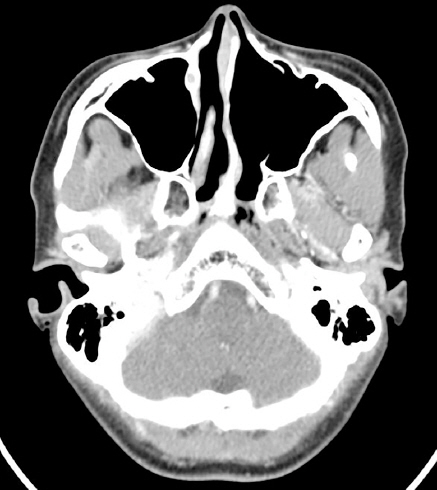
수술 후 12개월동안 외래 추적관찰을 진행 중으로 내시경 검진(Fig. 1C) 및 부비동 전산화단층촬영상(Fig. 4) 재발 및 합병증은 발생하지 않았으며, 좌측 코 막힘 증상은 호전되었음을 보고하였다.
고찰
근상피종은 주로 대타액선 혹은 때때로 부타액선에 발생하는 종양으로, 타액선 외 침범은 더욱 드물게 있으며, 그 중 비강 침범, 특히 비중격에서 기원한 경우는 더욱 드물게 보고될 정도로 희귀한 질환이다.1-13)
무통성, 무증상의 종물로 우연히 발견되는 경우도 있으나, 본 증례와 같이 일측성 코막힘을 호소하여 발견되는 경우 혹은 비출혈을 호소하는 경우도 보고되고 있다. 성비, 나이, 외상 유무와의 연관성은 명확히 밝혀진 바가 없다.11) 전산화단층촬영상 근상피종을 진단할 만한 특징적인 소견은 뚜렷하지 않지만, 치료 방침 및 절제 범위 결정에 도움을 줄 수 있다.
근상피종의 확진은 광학현미경상의 조직학적 소견을 통해 이루어지며, 종양은 고형, 점액양, 혹은 그물형의 성장 패턴을 보이고, 세포 형태는 방추형이 가장 흔하게 발견되며 이 외에도 형질세포양, 상피양, 투명세포형으로 구분되지만, 성장패턴이나 세포 형태가 예후에 영향을 미치진 않는 것으로 알려져 있다.11,14) 관세포형성이(ductal formation) 적다는 점이, 다형성 선종을 비롯한 다른 타액선 기원 종양들과의 차이가 되며, 비록 본 증례에서는 시행하지 않았지만 면역조직학적염색을 이용하면 감별 진단에 추가적인 도움을 받을 수 있다. 면역조직학적염색상 cytokeratin, S-100이 주요 표지자로 양성 반응을 보이고, calponin, smooth muscle actin, myosin, vimentin, glial fibrillary acidic protein (GFAP), carcinoembryonic antigen 등에도 양성 반응을 보이며, 이는 평활근종, 편평상피세포암을 배제하는데 도움이 되는 소견이고, 근상피종이 주위와 경계가 분명하며 주변조직침습을 하지 않는 것과 달리, 근상피암종의 경우 침윤성 변연과 함께 주변조직침습이 흔하며, 조직학적으론 조직 괴사, 혈관 및 신경주위침윤이 흔하게 관찰되는 것을 통해 감별할 수 있다.14,15)
치료는 외과적 완절절제가 원칙이며, 종양이 주위 조직들과 분명한 경계를 가지는 특징이 있어 완전 절제 후 국소 재발은 드문 편이고, 방사선치료 혹은 항암치료의 경우 근상피세포의 방사선 및 항암 감수성이 낮은 것으로 알려져 있어 일반적으로 시행되지는 않는다.8,12,14,15) 본 증례에서는 내시경부비동수술을 통한 종양완전적출술 시행 후 12개월동안 외래 추적관찰을 하였으며, 현재까지 재발 및 합병증은 발생하지 않고 있다.
최근 들어 비강 침범을 비롯한 근상피종의 타액선 외 침범의 보고가 점진적으로 증가하고 있으나, 이전까지 근상피종이 과소 진단 되었을 가능성을 배제할 수 없을 것으로 보이고, 병원 접근성의 증가와 면역조직학적 염색을 비롯한 병리학적 진단 기술의 발달 등이 최근의 진단에 도움을 주었을 가능성이 높을 것으로 보이며, 따라서 앞으로도 근상피종의 타액선 외 침범의 보고는 더욱 높아질 것으로 보인다. 그러므로 본 증례에서와 같이 일측성 비폐색과 종물을 주소로 내원한 환자에서, 내시경 검진 및 영상 검사상 혈관종, 기질화 혈종, 반전성 유두종과 같이 진단 특이적인 소견이 관찰되지 않는 경우, 근상피종과 같은 타액선 종양 역시 감별진단으로 고려되어야 할 것이다.
