수술 중 근적외선 자가형광으로 확인된 갑상선 내부의 부갑상선 선종 1예
A Case of Intra-thyroidal Parathyroid Adenoma Confirmed by Intraoperative Near-infrared Autofluorescence
Article information
Abstract
= Abstract =
In general, the anatomical location and number of parathyroid glands are well known, but they are often found in a variety of locations, making it difficult to find parathyroid glands during surgery. Besides Intra-thyroidal parathyroid adenoma is extremely rare case, and it is harder to identify in surgery. We encountered a 51-year-old patient with a thyroid nodule. The results of the additional blood test and the Tc-99m MIBI were combined to determine that the left lower lobe parathyroid adenoma was highly likely. This patient was treated with left thyroid lobectomy with parathyroid identification using Near-infrared (NIR) imaging. Afterwards, the biopsy confirmed that it was a parathyroid adenoma, and has since been monitored through outpatient observation without any problem. We present this rare case with a review of related literatures.
서론
일차성 부갑상선 기능항진증은 칼슘, 인산 및 골 대사에 이상을 초래하는 질환으로 생화학 검사의 발달에 따라 혈청 칼슘 측정과 부갑상선 호르몬의 측정이 간편해지면서 비교적 쉽게 진단이 진단 된다.1) 약 85%의 선종과 약 15%의 부갑상선 증식증, 그리고 약 1% 정도의 부갑상선 암종으로 구성된다.2)
부갑상선 선종의 진단과 수술적 치료의 기준은 비교적 잘 알려져 있지만, 수술로 제거하려는 기능이 항진된 부갑상선이 어디에 있는지 수술 전에 미리 알아보기 위한 술 전 국소화 검사에 대해서는 다양한 의견들이 있다. 임상적으로 가장 중요한 영상 검사는 일반적으로 부갑상선 스캔(Tc-99m MIBI) 검사가 흔히 이용되며, 민감도가 약 90%로 단일 검사로는 가장 정확하다고 알려져 있다.3)
일반적으로 부갑상선의 해부학적 위치와 개수는 잘 알려져 있지만, 발생학적으로 다양한 위치에서 발견되어 수술 중에 부갑상선을 쉽게 찾지 못하는 경우가 있다. 특히 드물지만 부갑상선이 갑상선 실질 내에 파묻혀 있다면 그 중 부갑상선 선종이 갑상선 내에 발생하는 경우는 전체의 1.4 - 4%에 불과하다고 알려져 있다.4) 수술 중에 수술 전 국소화 검사를 통해 부갑상선이 있을 것으로 예측되는 위치에서도 찾을 수 없는 곤란한 상황을 경험할 수도 있을 것이다.
최근에는 근적외선 자가형광(Near infrared autofluore- scence, NIR AF)을 통해 수술 중에 부갑상선을 확인할 수 있는 기술이 소개되었고, 근적외선의 조직 투과성을 이용하면 조기 발견도 가능하여 주변 조직의 박리를 최소한으로 줄여서 안전하게 부갑상선을 찾을 수도 있다.5)
저자들은 부갑상선이 드물게 갑상선 내에 위치하여 수술 중에 육안으로 찾기 어려웠던 부갑상선 선종 1례에서 근적외선 자가형광 영상을 이용하여 쉽고 정확하게 국소화를 할 수 있었던 경험을 문헌고찰과 함께 보고하는 바이다.
증례
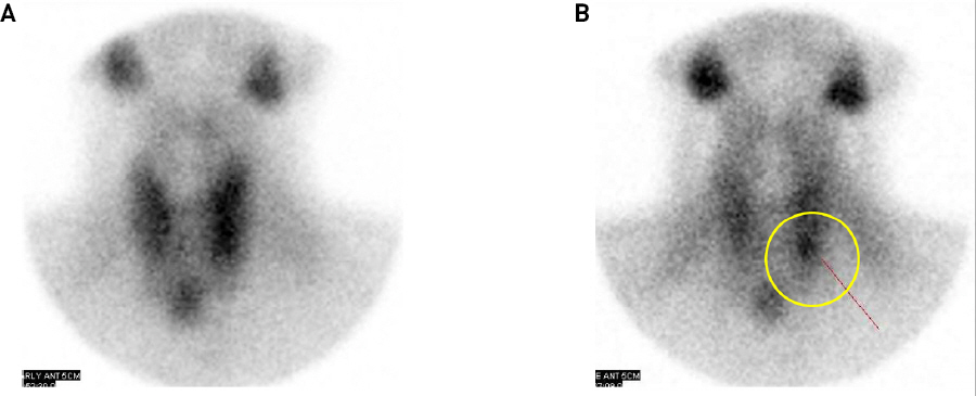
51세 여자가 타 병원에서 시행한 경부 초음파(Fig. 1)상 갑상선 내 관찰되는 결절에 대해 추가적 검사가 필요하여 본원 이비인후과 외래로 내원하였다. 타 병원에서 시행한 세침흡인검사 상 좌측 갑상선에서의 0.76cm 결절은 Atypia of undetermined significance (AUS)로 확인되었으며 본원에서 중심부바늘생검을 추가로 시행했다. 그 결과 부갑상선 선종과 증식 모두 가능성이 있었고, 부갑상선 호르몬 수치를 추가로 함께 확인하여 감별하기로 했다. 부갑상선 스캔(Tc-99m MIBI)에서는 좌측 하엽의 부갑상선 선종을 의심할 수 있는 소견이 확인되었다(Fig. 2).

Preoperative ultrasonography (USG) findings. (A) Transverse view shows 0.6cm sized mass (arrow) in left lobe of thyroid. The ultrasonic feature of the mass is heterogeneously hypoechoic, relatively well demarcated, and close to thyroid capsule. (B) Longitudinal view shows that the mass (arrow head) was located in the lower portion in left thyroid.

Preoperative 99mTc-sestamibi scan for localization. (A) Early phase. (B) Delayed phase shows high uptake in circled area, suspicious for parathyroid adenoma.
수술 1개월 전 혈액검사 상에서는 부갑상선호르몬 59.78 pg/ml (참고치 15-65), 칼슘 10.3mg/dL (참고치 8.0-10.0), 이온화 칼슘 1.34mmol/L (참고치 1.09-1.30)로 경도의 고칼슘혈증 소견이 확인되었으며 경부 이학적 검사상 비인강과 인후두에는 특이 소견이 없었고, 경부 촉진에서도 종물은 만져지지 않았다. 이상 검사 결과들을 종합하여 좌측 하엽 부갑상선 선종의 가능성이 높다고 판단했지만 조직학적 확진을 위해 환자와 상의 하에 좌측 갑상선 절제술을 계획하였으며 종양의 범위를 확인하기 위하여 시행한 경부 전산화단층촬영(Computed tomography, CT)에서 저명한 전이 소견은 관찰되지 않았다.
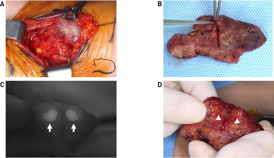
수술 중 갑상선을 외측화 시키면서 좌측 상엽의 부갑상선 위치를 예상하려 했지만 육안적으로는 확인이 힘들었으며 NIR AF를 통해 확인된 부분에서 부갑상선을 찾아낼 수 있었다(Fig. 3). 마찬가지로 하엽의 부갑상선을 찾고 동결절편 검사를 보내 부갑상선의 선종임을 확인했다. 좌측 갑상선 절제 직후, 20분 후, 30분 후 각각 혈액검사를 시행했으며 또한 갑상선을 절제한 후 선종이 의심되는 곳에 다시 절개를 가하고 NIR AF로 재확인하였다(Fig. 4).

Intraoperative findings. (A) Intra-thyroidal mass suspicious for parathyroid adenoma was pointed by surgical instrument. (B) Near infrared (NIR) imaging shows pointed area glowing brighter than background tissue (arrow). This lesion is highly suspicious for parathyroid tissue.

Gross specimen findings. Intra-thyroidal parathyroid adenoma was identified by additional incision in the dissected left thyroid gland. (A) There were no other structures suspicious for parathyroid gland in surgical field. (B) The suspicious parathyroid tissue was encapsulated by thyroid capsule, divided into two parts. (C) NIR imaging shows two parts of mass with bright white color (arrow). (D) Parathyroid tissue buried in thyroid gland was dissected (arrow head).
수술 중 시행했던 혈액검사는 갑상선 절제 20분 후 검사결과에서 부갑상선호르몬 10.38pg/ml (참고치 15-65), 이온화 칼슘 1.08mmol/L (참고치 1.09-1.30)로 술 전 시행했던 검사에 비해 부갑상선호르몬 기준 50%이상 감소한 것을 확인 할 수 있었다. 이후 입원 중 술 후 3시간, 술 후 1일차, 3일차 총 3차례의 추가 혈액검사를 시행했으며 각각 모든 검사에서 칼슘, 이온화 칼슘, 부갑상선호르몬 수치가 정상범위 내인 것을 확인하였다(Table 1).
최종 조직검사결과 크기 0.7 x 0.7 x 0.7cm의 부갑상선 선종이 확인되었다(Fig. 5). 수술 후 환자는 특별한 후유증 없이 퇴원하였고 현재 외래에서 3년 6개월 동안 추적관찰 중이다.
고찰
부갑상선의 종양은 약 85%의 선종과 약 15%의 부갑상선 증식증, 약 1% 이하의 부갑상선 암종으로 구성되며,2) 부갑상선 선종이 부갑상선 기능항진을 동반할 때에는 대부분의 환자들이 술 전에 원인을 모르는 장기간의 근골격계 증상, 신장증상, 소화기계 증상 외에도 늑골종물, 손저림, 음성변화, 무증상과 같은 다양한 증세를 주소로 내원하게 된다. 대상환자들은 부갑상선 기능항진증으로 진단되기까지 원인을 모르는 이러한 비특이적인 증세로 인하여 장기간 치료를 받았으며 대부분의 환자들은 병원을 내원 후에도 정형외과, 비뇨기과 등의 여러 과에서 진료를 받으며 부갑상선 선종으로 진단받기까지 짧게는 1달에서 길게는 9개월까지의(평균 2.7개월) 기간이 걸린다.2)
술 전 부갑상선 스캔(Tc-99m MIBI)은 부갑상선 병변을 국소화 하는데 도움이 될 수 있지만 일부 부갑상선 병변은 그 위치, 작은 크기, 혈청 칼슘과 부갑상선 호르몬 수준의 상승 등으로 도움이 되지 못할 수도 있다.3) 그 외에도 술 전 부갑상선의 위치를 국소화하기 위해 경부 초음파, 전산화단층촬영(CT)이 도움이 될 수 있다.6,7)
부갑상선 스캔(Tc-99m MIBI)은 민감도가 88%라고 보고되었으며,7) 단일 영상검사로는 가장 정확하다고 알려져 있다.3) 경부 초음파의 경우 방사능 노출이 없으며, 타 검사에 비해 저렴하고 간편하다는 장점이 있다. 일반적으로 부갑상선 선종의 경우 초음파에서 저에코의 균일하고 경계가 비교적 뚜렷한 구조가 관찰된다. 하지만 정상 부갑상선은 초음파에서 발견하기 어렵다. 술 전에 단독 영상으로 시행되는 경우는 거의 없어 보조적으로 시행된다. 전산화단층촬영은 검사 시간이 짧고, 작거나 위치가 비특이적인 경우에도 잘 확인되어 단일 부갑상선 선종에서의 민감도가 94%라는 장점이 있다. 그러나 부갑상선 스캔에 비해서 57배의 방사능 노출이 발생하여 이론적으로는 갑상선 암종의 위험성을 높일 수 있다는 단점이 있다.8)
수술 중 부갑상선을 국소화 시키는 방법으로는 전통적으로 갑상선을 노출시킨 후 가능하면 무혈 시야에서 중갑상정맥을 찾아 결찰하고 갑상선엽을 전내측으로 견인하여 반회후두신경 및 하갑상동맥을 확인한다. 하부갑상선은 발행학적인 특성으로 위치의 분포가 상부갑상선의 비해 다양하다. 약 50%에서 갑상선엽 하극(lower pole)의 측후방 부위 0.5 cm 이내의 하갑상동맥 분지 부근에 위치하며, 12.8%에서는 1 cm 이내에, 약 10%에서는 종격동에 위치한다. 본 증례와 같이 부갑상선 선종이 갑상선 내에 위치하는 경우는 전체의 1.4 - 4%에 불과하여 비교적 드문 경우이다.4)
수술 중에 육안으로 부갑상선의 저명한 소견이 관찰되면 부갑상선만 안전하게 절제할 수도 있겠지만, 본 증례와 같이 갑상선 조직 내에 파묻혀 있는 경우라면 동측의 갑상선을 절제하는 것이 일반적이다.9,10) 수술 중에 부갑상선을 찾는 다양한 방법들을 적용하여 평가해 보았지만, 최근에는 부갑상선 조직이 근적외선 영역에서 형광현상을 보이는 것을 알게 되었고, 이를 영상으로 구현하는 기술이 소개되었다. 그 원리를 아직은 명확하게 알 수는 없지만 어떠한 조영 물질을 주입하지 않아도 되기 때문에 자가형광(autofluorescence)이라고도 한다.11)
부갑상선의 자가 형광 현상에서 적용되는 빛의 스펙트럼은 근적외선(700-900 nanometer)인데, 이 파장대는 조직내의 물과 혈색소에 흡수되는 정도가 낮아 조직을 수 밀리미터까지 침투할 수 있다는 특성이 있다. 그래서 부갑상선이 주변의 지방조직이나 결체조직에 파 묻혀 아직 노출되지 않은 상태에서도 조기 발견이 가능하다는 보고가 있다.12) 본 증례도 갑상선 조직 내에 부갑상선 선종이 위치하고 있어서 수술 영역과 갑상선 검체의 표면에서 부갑상선이 육안으로 보이지는 않았지만, 그 위치를 정확하고 쉽게 확인할 수 있었기에 매우 유용하였다. 이는 수술 시간을 감소시킬 수 있는 중요한 요소였다.
또한 수술 중 부갑상선호르몬의 혈중 농도를 시간대별로 측정하는 것(intraoperative parathyroid hormone monitoring)이 남은 부갑상선 조직의 기능을 평가하고, 수술적 치료가 잘 되었는지를 평가하는 전통적인 방법으로 본 증례에서도 함께 시행하였다.
부갑상선 질환의 수술적 치료를 고려할 때는 수술 전, 수술 중 부갑상선을 국소화 하는 것이 중요하며 알려진 다양한 방법들에 더해 최근에는 자가형광 비디오 모니터링 또한 유용한 방법으로 생각된다. 갑상선 조직 내에 있는 경우 동측의 갑상선을 함께 절제하는 것이 일반적이지만9,10) 본 증례는 NIR AF를 이용하여 국소화 할 수 있었다.
이를 활용하여 부갑상선 선종을 포함한 갑상선 부분절제술을 시행하고, 수술 중 부갑상선호르몬 혈중 농도 측정으로 수술적 치료의 완전성에 대한 모니터링을 한다면 불필요한 갑상선 수술의 범위를 줄일 수 있을 것으로 판단된다. 이에 본 증례는 향후 더 좋은 수술적 치료의 가능성을 제시할 수 있는 증례라 판단되어 문헌고찰과 함께 보고하는 바이다.

