전신에 새로이 발생한 다발성 기저세포암 1예
Reoccurred Multiple Basal Cell Carcinomas: A Case Report
Article information
Abstract
= Abstract =
Basal cell carcinoma (BCC) is the most common skin cancer. Ultraviolet radiation exposure and genetic predisposition are known to be the most important etiological factors. Multiple BCC is often associated with genetic familial conditions such as BCC syndrome, basal cell nevus syndrome. We present a case of 54-year-old female who had multiple BCC that had reoccurred. She was completely cured after receiving radio-chemotherapy for leukemia 16 years ago. She had multiple lesions (scalp, left thigh, right popliteal fossa, and right buttock), and had underwent wide excisions of all lesions. All biopsies revealed BCC. Six years later, she had also multiple lesions; left forehead, frontal vertex scalp, parietal vertex scalp, right occipital scalp, and lower abdomen. We performed wide excision. Histopathological examination revealed BCC. She had no signs of any BCC associated syndrome. We report a rare case of nonsyndromic multiple BCC that reoccurred at the new site.
서론
기저세포암(Basal cell carcinoma, BCC)은 가장 흔한 피부암으로 기저세포암으로 인한 사망률은 높지 않지만 이환율이 높아 의료서비스에 부담이 커지고 있다.1) 자외선 노출 및 유전적 소인이 기저세포암의 가장 유의한 원인으로 알려져 있으며 그 밖에 Fitzpatrick 피부 유형 I & II, 고령, 남성, 유해물질 노출, 지방 과다 섭취 등이 병인학적 요인으로 알려져 있다.2,3) 다발성 기저세포암은 보통 기저세포암 증후군과 같은 유전적 질환과 연관되어 나타난다.2) 저자들은 이러한 증후군의 특징을 나타내지 않는 다발성 기저세포암이 새로이 발생한 환자를 경험하여 이에 대한 문헌고찰과 함께 본 증례를 보고하고자 한다.
증례
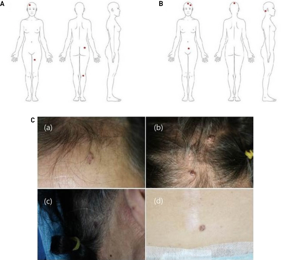
54세 여자 환자가 전신의 다발성 피부병변을 주소로 내원하였다. 환자는 내원 16년 전 백혈병으로 전신 방사선 치료와 항암화학요법을 받은 후 완치되었으며 다른 과거력은 없었다. 피부암의 가족력은 없었다. 환자는 내원 6년전에 본원에서 전신의 다발성 기저세포암으로 수술적 치료를 받았었다. 처음 내원 당시 3년전부터 발생한 전신의 다발성 피부병변이 있었으며, 병변은 전두부 두피의 1×2 cm의 흑색 가피 유사 병변과 왼쪽 허벅지(0.5x1 cm), 오른쪽 슬와(0.5x0.5 cm) 및 오른쪽 엉덩이(1x1 cm)의 압통이 없는 흑색 결절이었다(Fig. 1A). 이에 대해 본원 피부과에서 펀치생검을 시행한 결과 기저세포암 진단을 받고 수술적 치료를 위해 성형외과로 의뢰되었다. 저자들은 모든 병변을 동결절편 조직검사와 함께 광범위 절제술을 시행하였다. 모든 병변의 병리조직검사 결과는 기저세포암으로 보고되었다. 수술 후 뇌 자기공명 영상(MRI)과 전신의 양전자 방출 단층촬영(PET-CT)을 시행한 결과 원격전이 소견은 없었다. 이후 약 6년이 지난 뒤 내원한 환자에서 왼쪽 이마의 0.5×0.5 cm 흑색 유색 황반 병변, 전두부 두피의 0.7×0.5 cm 흑색 유색 구진과 정수리 두피의 1.5×0.3 cm 회백색 사마귀양 종괴, 우측 후두부 두피의 0.5×0.5 cm 흑색 유색 황반 병변, 그리고 하복부의 1×1 cm 흑색 색소 결절 이 관찰되었다(Figs. 1B, 1C). 이 병변들은 지난 수술 시 병변들과 모두 다른 위치에 발생하였다. 저자들은 이전과 마찬가지로 모든 병변을 동결절편 조직검사와 함께 광범위 절제술을 시행하였으며 모든 병변의 병리조직검사 결과는 기저세포암으로 보고되었다. 환자가 유전적 검사를 포함한 모든 추가 검사를 거부하여 추가적인 평가는 불가능했다.

(A) 1 × 2 cm black eschar-like lesion on the scalp, 0.5 × 0.5 cm left thigh, right popliteal fossa (0.5x0.5 cm) and right hip (1x1 cm) no tender black nodule. (B, C) (a) 0.5×0.5cm black pigmented macular lesion of left forehead, (b) 0.7×0.5cm black pigmented papule of frontal scalp and 1.5×0.3cm grayish verrucous soft mass of parietal scalp, (c) 0.5×0.5cm black pigmented macular lesion of right occipital scalp area, (d) 1×1 cm black pigmented nodule of the lower abdomen.
고찰
기저세포암은 사람에게서 발생하는 가장 흔한 암으로 보통 얼굴과 목에 단일 병변으로 나타난다.3) 기저세포암은 유전적 소인과 외부 환경적 요인으로 인해 복합적으로 발병하며 발생기전 중 가장 유의한 위험요소는 자외선 노출로 인한 종양억제유전자의 변이로 알려져 있다. 그리고 방사선, 비소 등에 노출되는 경우 조절유전자의 변이를 일으키고 면역감시체계의 변화를 일으켜 기저세포암을 유발시키는 것으로 알려져 있다. Gorlin 증후군, Rombo 증후군, 일측성 기저세포 모반 증후군 및 Bazex 증후군은 다발성 기저세포암이 나타나는 유전적 질환이다.2) 그러나 이러한 유전적인 소인이 없이 발생한 다발성 기저세포암에 대한 위험인자는 아직 잘 알려져 있지 않다. Verkouteren의 연구 결과에 따르면 처음 단일 BCC가 발생했을 때 보다, 한 개 이상의 BCCs가 발생했을 때 metachronous BCCs가 발생할 확률이 2.5배 높은 것으로 알려져 있다.4) 기존의 연구결과들에서는 다발성 기저세포암의 예측인자로 젊은 연령, 피상적 기저세포암(superfi- cial BCC subtype), 빨간 머리카락 표현형, 몸통이나 상지에 발생한 기저세포암, 남성으로 보고하였으며,2) 다발성 기저세포암은 자외선 노출 등의 외부 환경적 요인보다는 환자의 유전적 감수성이 더 큰 영향을 미치는 것으로 나타났다.4,5) 면역억제 상태는 다양한 피부암을 유발하는 요인으로 알려져 있는데 만성 림프구성 백혈병으로 인한 면역체계 손상에 따른 2차 악성종양으로 편평상피세포암(Squamous cell carcinoma)과 기저세포암이 발생할 수 있다.6,7) 면역억제 상태와 관련된 비흑색종 피부암은 특발성 피부암과 달리 편평상피세포암이 기저세포암보다 더 많이 나타난다.8) 방사선의 노출도 비흑색종 피부암의 위험인자로 알려져 있다. 히로시마와 나가사키 원폭 피해자와 두피백선으로 방사선 치료를 받은 환자들에서 비흑색종 피부암의 높은 발병률을 보였으며, 그 중 각각 50 %와 98 %가 기저세포암으로 나타났다.8,9) Watt 등의 연구에서는 피부에 35 Gy이상의 선량으로 방사선 치료를 받은 환자의 경우 방사선 치료를 받지 않은 환자에 비해 기저세포암의 발병위험이 약 40배가 높은 것으로 나타났으며, 1 Gy이상의 피부 방사선량은 기저세포암의 발병 위험의 증가와 관련이 있다고 하였다.10) 본 증례의 환자는 유전적 질환의 증상이나 가족력이 없이 다발성 기저세포암이 발생한 환자였으며 처음 발병 후 6년 뒤 다른 위치에 다발성 기저세포암이 새로이 발생하였다. 광범위 절제술을 시행하였으며 미용적인 결과와 종양의 치료적 측면을 모두 고려하여 안전 절제연의 거리는 0.3-0.5 cm로 하였으며 수술 중 동결절편 생검을 통해 절제연을 확인하였다. 환자는 첫 수술적 치료로 4개의 기저세포암을 제거 받았고 두번째 수술적 치료로 5개의 기저세포암을 제거 받았다. 저자들은 이 환자가 이전에 백혈병이 있었으며 백혈병의 치료를 위해 시행 받았던 전신 방사선 치료가 다발성 기저세포암의 원인이 되었을 것으로 생각한다. 따라서 본 증례는 다발성 기저세포암이 다른 위치에 새로이 발생한 매우 드문 사례로 사료되어 이에 보고하는 바이다.
Notes
Conflict of interest
No potential conflict of interest relevant to this article was reported.
Ethical approval
The study was approved by the Institutional Review Board of Nowon Eulji Medical Center, Eulji University (IRB No. 2022-09-004) and performed in accordance with the principles of the Declaration of Helsinki. Written informed consent was obtained.
Patient consent
The patient provided written informed consent for the publication and the use of their images.